


Abstract
Kienbock’s disease is a condition characterized by avascular necrosis of the lunate bone. It is also known as lunatomalacia or aseptic and ischemic necrosis of the lunate. In a patient diagnosed with COPD who had uncontrolled use of oral corticosteroids, Kienböck’s disease developed after prolonged corticosteroid use. The patient had no history of trauma or other known risk factors associated with Kienböck’s disease. This case suggests that corticosteroid use may be an important risk factor among the systemic risk factors for Kienböck’s disease.
Cover Letter
Summary
Kienböck's disease is a condition characterized by avascular necrosis of the lunate bone. This disease is also known as lunatomalacia and aseptic or ischemic necrosis of the lunate bone . Patients with COPD diagnosis who used unchecked oral corticosteroids developed Kienböck's disease after prolonged corticosteroid use. The patient had no history of trauma or other risk factors known to be associated with Kienböck's disease. This case suggests that among the Systemic risk Factors of Kienböck's disease, corticosteroid use may be a prominent risk factor.
Introduction:
Kienböck's disease is a condition characterized by avascular necrosis of the lunate bone . And it is manifested by pain, swelling and stiffness in the dorsomedial part of the wrist . As Risk factors, it has been associated with genetic, anatomical, vascular, metabolic and systemic factors. However, the exact etiological factors have not yet been fully clarified. In the clinical case that we will present, it was mentioned that Kienböck's disease was detected in a 73-year-old male patient with a systemic risk factor who was treated with a diagnosis of COPD exacerbation.
Clinical case:
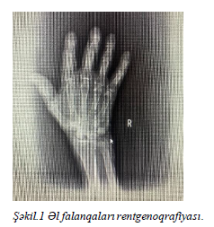
A 73-year-old male patient came to our clinic with complaints of shortness of breath, shortness of breath, neuropathic pain in the limbs, weakness. The patient has reported suffering from COPD for many years and has been taking oral steroids for a long time due to this. The objective examination shows pale skin and mucous membranes, lipomas in various parts of the body, and a discharge wound in the left right area. Hallux-valgus symptom is observed on the legs. The anatomical structure of the toes is impaired. In auscultation, dry whistling breathing is heard over the lungs. With 3 liters of oxygen from the nose, SpO2 is - 94%. In laboratory analyzes, it was measured as WBC - 11.41 x 10^3/µL; HGB -14.7 g/dL; MCV-84 fL; PLT-179 x 10^3 ALT - 27.4 u/l, AST - 15.5 U/l, CRP - 18.66 mg/l. Treatment of COPD exacerbation to the patient (inhalation with bronchiolitics and I for 5 days.V 40 mg steroids) and Ampicillin - sulbactam 4x1.5 gr was started due to the discharge wound in the dressing area. The patient had a history of Hallux-Valgus symptom, pain in the joints and a history of replaceable swellings in the joints and a runny wound, mainly consulted with a rheumatologist. As a result of clinical and laboratory evaluation, rheumatological disease was not considered. 3 days after the start of treatment, the patient began to improve dyspnea due to an exacerbation of COPD, bronchial spasm decreased, SpO2 in the room air was measured as 96%, discharge from the wound in the dressing area stopped. However, after 2 days, the patient had complaints of swelling and pain in the wrist area. In repeated laboratory evaluation, C - reactive protein (CRP) - 23.07 mg/L creatinine - 0.82 mg/dL was seen as urea (urea) - 62.3 mg/dL, Urea nitrogen (BUN) - 29.1 mg/dL. Consultation with the traumatologist, hand-wrist X-ray and ENMG planned hand phalanx X-ray ‘the intensity of the shading of the bones included in the examination area was sharply reduced in the periarticular areas. The subchondral bone rash of the interphalangeal joint surfaces of the right hand has become rough, the width of the joint clefts is limited. Osteophytes are traced at the edges of the articular surfaces in the phalanges’ (Fig.3). In enmg examination, nerve conduction could not be objectively assessed due to edematous and swollen hands of the patient on the right. On the exacerbation of the patient's pain complaints, the endocrinologist was consulted for a long time due to steroid intake, and in the foreground of the clinical evaluation of the patient thought Kienböck'S disease hand-wrist MRI was taken. At the same time, densitometry was planned and its result was evaluated as Osteopenia. Hand, hand-wrist MRI result " symptoms of osteoarthrosis. Severe fluid increase in the bed of the flexor tendons ( tenosynovitis). Os lunatum volume is slightly reduced, 6 mm in size T1 hypointens, T2 / FS hyperintens area (thought to be secondary to osteonecrosis - a symptom of Kienbock disease)’ reported. The treatment of Kienbock's disease was planned by a traumatologist. We do not have information about the dynamics of the patient after the control does not come.
Discussion
Kienböck's disease is a rare disease characterized by avascular necrosis of the lunate bone. This condition is usually manifested by pain in the wrist, restriction of movement, and swelling . Although the etiology of the disease has not been fully clarified, the role of trauma, genetic predisposition, vascular anomalies and systemic diseases is significant. In this case, prolonged steroid use by the patient has been recognized as one of the main risk factors in the development of Kienböck's disease. Corticosteroids can affect the vascular system, causing decreased blood flow and metabolic disorders in bone tissue. This can lead to the development of avascular necrosis in the lunate bone. The absence of trauma or other known risk factors in our patient supports that corticosteroid use plays an important role in the etiology of the disease. Radiological evaluation, especially MRI, is one of the main tools in the diagnosis of Kienböck's disease . In this case, an MRI scan confirmed the symptoms of osteonecrosis in the lunate bone. Other problems, such as the systemic hallux-valgus symptom and osteopenia, observed in the patient, point to a general skeletal weakness, further emphasizing the role of corticosteroids in these disorders. This clinical case confirms the multifactorial nature of Kienböck's disease and that corticosteroid use is a potential risk factor for this disease. Also, timely recognition of such cases and the correct implementation of treatment plans can improve the patient's quality of life.
The result
Kienböck's disease is a difficult clinical case in terms of diagnosis and treatment. In particular, this disease should be considered in patients with systemic risk factors, such as steroid use. Although non-surgical treatment can be effective in the early stages of the disease, surgical intervention is required in advanced cases. This case suggests that long-term corticosteroid use may be a major risk factor in the development of Kienböck's disease, emphasizing the need for more large-scale studies in the future. Doctors should pay close attention to complaints of wrist pain and restriction of movement, especially in patients who use steroids for a long time, and plan appropriate examinations in good time.
Figures
Keywords
References
Mənbələr:
1. Allan CH, Joshi A, Lichtman DM. J Am Acad Orthop Surg. 2001 Mar-Apr;9(2):128-36. PMID: 11281633. 2. Chojnowski K, Opiełka M, Piotrowicz M, Sobocki BK, Napora J, Dąbrowski F, Piotrowski M, Mazurek T. Recent Advances in Assessment and Treatment in Kienböck's Disease. J Clin Med. 2022 Jan 27;11(3):664. doi: 10.3390/jcm11030664. PMID: 35160115; PMCID: PMC8836398. 3. Camus EJ, Van Overstraeten L. Kienböck's disease in 2021. Orthop Traumatol Surg Res. 2022 Feb;108(1S):103161. doi: 10.1016/j.otsr.2021.103161. Epub 2021 Nov 30. PMID: 34861414.
4. Kahn SJ, Sherry DD. Kienbock’s Disease — Avascular Necrosis of the Carpal Lunate Bone — in a 7-Year-Old Girl With Dermatomyositis. Clinical Pediatrics. 1994;33(12):752-754. doi:10.1177/000992289403301210
5. Bae JY, Shin YH, Choi SW, Moon SH, Park HS, Kim JK. A novel classification of Kienbock's disease based on magnetic resonance imaging. Int Orthop. 2023 Aug;47(8):2023-2030. doi: 10.1007/s00264-023-05861-3. Epub 2023 Jun 10. PMID: 37300563.
6. Bartelmann U, Kalb K, Schmitt R, Fröhner S. Radiologische Diagnostik der Lunatumnekrose [Radiologic diagnosis of lunate necrosis]. Handchir Mikrochir Plast Chir. 2001 Nov;33(6):365-78. German. doi: 10.1055/s-2001-19454. PMID:11917675.
Article Info:
Publication history
Published: 25.Feb.2025
Copyright
© 2022-2025 Azerbaijan İnternal Medicine Society. Published by "Uptodate In Medicine" health sciences publishing. All rights reserved.Related Articles
Viewed: 363